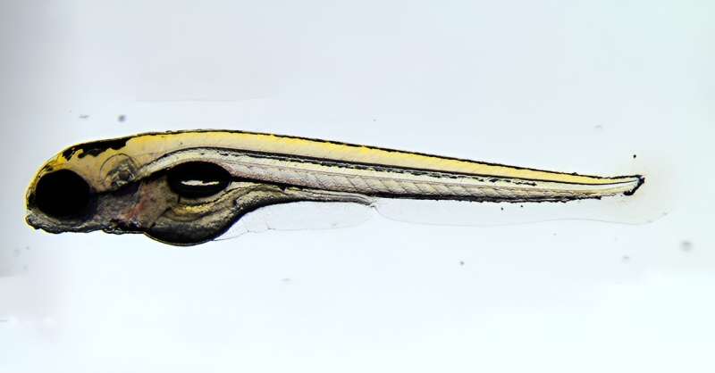
Researchers create genetic atlas detailing early stages of zebrafish development

Researchers at the National Institutes of Health have published an atlas of zebrafish development, detailing the gene expression programs that are activated within nearly every cell type during the first five days of development, a period in which embryos mature from a single cell into distinct cell types. These diverse cells become tissues and organs that form juvenile fish capable of swimming and looking for food. The findings are published in Developmental Cell.
“Perhaps surprisingly, tiny zebrafish provide us with significant insight into human development and disease. Many of the gene expression programs that direct embryonic growth are similar across fish, people, and other animals,” said Christopher McBain, Ph.D., scientific director of the Eunice Kennedy Shriver National Institute of Child Health and Human Development (NICHD), which conducted the work.
“Since zebrafish are visibly transparent, fertilize eggs externally, and are easy to study genetically, they represent a unique and effective way to model human disease.”
The process of embryonic development is orchestrated by instructions in DNA that direct different programs of gene expression within individual cells, which give different cell types their unique functional characteristics. To create the atlas, the study team used a method called single-cell RNA sequencing to identify gene expression programs over the course of five days, with samples taken every two to 12 hours.
The resulting atlas follows nearly 490,000 cells continuously over 120 hours after fertilization, with an average of 8,621 transcripts and 1,745 genes detected per cell. The study team then sorted these data among known cell types and cell states during development.
To highlight the atlas’ utility, the team focused on the development of understudied cells, including intestinal cells called BEST4+ cells, which are linked to gastrointestinal diseases and cancer in people. Little is known about how these cells develop because they are absent in other common model organisms, such as mice.
By using the atlas, the team computationally predicted the full developmental program of BEST4+ cells, including signals that initiate the cells’ development and transcription factors that carry out the process. These findings can be evaluated in model organisms or clinical samples to better understand the role of BEST4+ cells in human disease.
“Our atlas on early zebrafish development is an extremely thorough resource that describes the expression program of hundreds of cell types across 62 developmental stages,” said senior author Jeffrey A. Farrell, Ph.D., an Earl Stadtman Investigator and head of NICHD’s Unit on Cell Specification and Differentiation.

“From this atlas, we made discoveries about understudied cells, including intestinal cells involved in human diseases, smooth muscle that surrounds the intestine, and cells that surround blood vessels. There are many more advances waiting to be uncovered, and we look forward to seeing what the research community can do with our open-source atlas.”
The atlas is publicly accessible to the broader research community at https://daniocell.nichd.nih.gov. In addition to browsing the data online through the website, data may also be downloaded in additional formats for reanalysis. A timelapse of early zebrafish development is available for viewing, as well as microscopy images related to the study. To learn more about the NIH Zebrafish Facility, visit the NIH Virtual Tour.
More information:
Abhinav Sur et al, Single-cell analysis of shared signatures and transcriptional diversity during zebrafish development, Developmental Cell (2023). DOI: 10.1016/j.devcel.2023.11.001
Citation:
Researchers create genetic atlas detailing early stages of zebrafish development (2023, December 18)
retrieved 19 December 2023
from https://medicalxpress.com/news/2023-12-genetic-atlas-early-stages-zebrafish.html
This document is subject to copyright. Apart from any fair dealing for the purpose of private study or research, no
part may be reproduced without the written permission. The content is provided for information purposes only.


