Researchers make breakthrough in fighting a leading cause of fungal pneumonia

Scientists at Tulane University School of Medicine have developed a promising new model to study a pneumonia-causing fungus that has been notoriously difficult to culture in a lab.
Researchers were able to use precision-cut slices of lung tissue to study Pneumocystis species, a fungus that causes Pneumocystis pneumonia in immunosuppressed patients and children.
This innovation overcomes a major hurdle in fungal research—the difficulty of growing this pathogen outside of a living lung—so scientists can more easily test new drugs to fight the infection. The fungus was recently listed among the top 19 fungal priority pathogens by the World Health Organization.
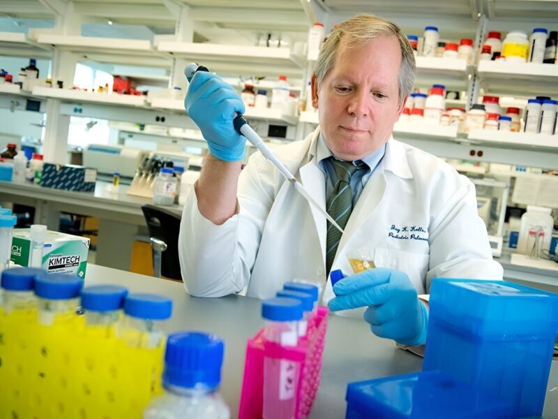
“Pneumocystis is likely the most common fungal pneumonia in children and attempts at culturing the organism have largely not been successful,” said corresponding author Dr. Jay Kolls, John W Deming Endowed Chair in Internal Medicine at Tulane. “Thus, we have not had new antibiotics in over 20 years as they have to be tested in experimental animal studies.”
The Tulane model utilizes precision-cut lung slices which retain the complexity and architecture of lung tissue, providing an environment that closely mimics conditions inside the lung. The results were published in mBio.
Researchers used tissue from mice to cultivate two forms of the Pneumocystis fungus—the troph and ascus—for up to 14 days. The viability testing and gene expression analysis they conducted showed the fungus survived over time in the model.
“This is the first time both the trophic and ascus forms of Pneumocystis have been maintained long-term outside a mammalian host,” Kolls said.
The researchers confirmed the model’s potential for in vitro drug testing. When treated with commonly used medications trimethoprim-sulfamethoxazole and echinocandins, the expression of Pneumocystis genes was reduced, indicating successful targeting of the fungus.
The Tulane technique reliably generates many uniform lung tissue samples for experimentation from a single lung, enabling high-capacity testing.
“With optimization, we believe precision lung slices could enable actual growth of Pneumocystis and become a powerful tool for developing new medications to treat this infection,” Kolls said. “This could significantly accelerate research on this pathogen.”
The study was led by Ferris T. Munyonho, a Tulane Biomedical Sciences graduate student and a recipient of a Fulbright Scholarship after receiving his Bachelors’ of Science from the University of Zimbabwe.
More information:
Ferris T. Munyonho et al, Precision-cut lung slices as an ex vivo model to study Pneumocystis murina survival and antimicrobial susceptibility, mBio (2023). DOI: 10.1128/mbio.01464-23
Citation:
Researchers make breakthrough in fighting a leading cause of fungal pneumonia (2023, December 28)
retrieved 29 December 2023
from https://medicalxpress.com/news/2023-12-breakthrough-fungal-pneumonia.html
This document is subject to copyright. Apart from any fair dealing for the purpose of private study or research, no
part may be reproduced without the written permission. The content is provided for information purposes only.


